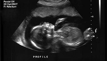
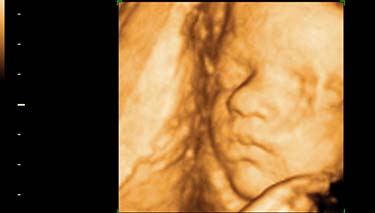
Most women will have at least one pregnancy ultrasound, either in very early stages of their pregnancy or at around 20 weeks of gestation. So even if you have never personally had one, you will probably have heard of pregnancy ultrasounds and aren t entirely unfamiliar with how they are done. Pregnancy ultrasounds are just one of the many screening tools available to assess that all is going well with a mother's pregnancy and the growth and development of her baby.
Other names for pregnancy ultrasounds are a pregnancy dating scan, commonly done within the first trimester, and the pregnancy anomaly or screening ultrasound. This is most commonly done at around 18-20 weeks of gestation. If the maternity care provider is concerned about the baby's growth in the third trimester, then an additional ultrasound at this stage may be recommended. Determining the baby's size, weight and growth as well as the amount of amniotic fluid present can provide a good, comprehensive assessment of the baby's status.
Sometimes during a third trimester ultrasound, findings reveal that the baby would be safer in a neonatal intensive care unit than to remain in the uterus. And despite the risks associated with prematurity these are not considered to be as significant to the baby's health if the pregnancy continued.
What is a pregnancy ultrasound?
This is when an ultrasound machine sends high-frequency sound waves through a mother's uterus of her vagina. These sound waves are higher than can be registered by the human ear, which is why pregnancy ultrasound is a quiet procedure. The sound waves are emitted from a small, vibrating crystal housed in a hand-held device known as a transducer. It is a painless procedure and will not hurt you or your baby. During pregnancy ultrasound, the sound waves which are created bounce off the baby and in turn are interpreted into an image on a screen. These images are occurring in real time; there is no delay between when the images are picked up and when they can be seen on the screen. So the baby can be seen moving around and kicking, as it actually is, rather than snapshots of still imagery.
Warm, translucent gel is first applied to the mother's abdomen. This helps to reduce the traction between the transducer and the skin and assists in transmitting the ultra sound waves with less interference so they can pass from the transducer through mother's body.
Hard tissues such as bone appear as white and soft tissues appear as grey. Amniotic fluid appears black because the sound waves just go through them as there are no firm tissues for them to bounce off. Each shade and its intensity are interpreted by a sonographer and provide different information.
What will I be able to see?
You will be able to see a lot of your baby, in terms of their physical shape and appearance all of their internal organs including their brain, heart, lungs, kidneys, liver and stomach; also their spine, their limbs, their genitals and their movements. You will be given a photograph of one or two of the ultrasound images to take home with you, but don t expect the scan to provide the same level of detail as a photograph. You may not get a video of your baby either. Some practices are not including this as an option due to the risk of litigation if complications are not detected. Speak with the individual sonography service to see what you can actually take home with you.
Will a pregnancy ultrasound hurt me or my baby?
Pregnancy ultrasound has been done routinely in Australia and New Zealand for the last thirty to forty years. There have been multiple research studies conducted over the years looking at the potential risks to mothers and their babies, but to date there is no consistent evidence that ultrasound is unsafe for either. Pregnancy ultrasound is also non invasive which means that it will be done through the skin on your abdomen or vaginally. An ultrasound is not a surgical procedure; there is no cutting into or around the skin. It does not use radiation, unlike an X-Ray, so it is considered much safer to use as a diagnostic tool during pregnancy.
Currently, the sonography industry is well monitored and regulated and the staff training very comprehensive. Due to its popularity, if concerns about risk factors ever do evolve, immediate changes to pregnancy ultrasound practice would occur.
Why should I have a pregnancy ultrasound?
There are many reasons for having a pregnancy ultrasound, but some of the most common are:
- To diagnose a viable pregnancy.
- To assess the gestational age of the baby.
- To check the baby's growth and development.
- To check how many babies are present in the mother's uterus.
- To see where the placenta is positioned.
- To do a nuchal fold measurement at the back of the baby's neck and to assess if there is an increased risk of the baby having Down Syndrome. This specific measurement needs to be conducted between 11 14 weeks.
- To assess for specific problems the baby may have, such as spina bifida.
- To assess the baby's gender; though this is not a primary reason for pregnancy ultrasound it is often determined at the 20 week screening scan.
- To assess the cause of pregnancy complications. This may be bleeding, a reduction in the baby's movements or if the mother has been experiencing pain.
- To detect whether there is an ectopic pregnancy. This is when the baby is growing outside of the mother's uterus, most commonly in one of the fallopian tubes.
- When an amniocentesis or chorion villus sampling test is being conducted, a pregnancy ultrasound is done at the same time. This is to ensure that the needle is inserted into the correct area and there is no risk to the baby.
- To check for uterine fibroids or ovarian cysts.
Where will I go for a pregnancy ultrasound?
Most large suburbs have radiography clinics which provide pregnancy ultrasound services. You may be referred to one of these, or alternately to your local maternity hospital radiography department, community health centre or a pregnancy specific/women's health sonography service.
The procedure generally takes around thirty minutes. If you have been booked for a vaginal ultrasound and then need to have an abdominal scan, you may be there for up to one hour. Try not to rush. You will want to enjoy your pregnancy ultrasound as much as possible and if you are stressed and anxious, you won t get everything out of it that you otherwise could.
When will I get the results of my pregnancy ultrasound?
Immediately, as the sonographer is doing the procedure. They will turn the screen around at some stage during your pregnancy ultrasound and point out to you various images on the screen. You may also go to a clinic which provides a separate screen to the one the sonographer needs, specifically for parents to look at.
If any concerns arise during the ultrasound, the sonographer will request a second opinion from a colleague. Sometimes this is not possible and can t happen at the same time which means there may be an unavoidable wait. Of course, this can be very worrying for parents, but many times, there is simply a variation of normal which just needs to be clarified.
The results of the scan will be sent to the referring health professional who recommended the scan and you will be informed of the results. Of course, complications can be detected during ultrasound which is one of the reasons why it is done in the first place. In this case you will be encouraged to speak with your maternity care provider who will be informed by the radiographer's assessment.
Who will do the pregnancy ultrasound?
Pregnancy ultrasounds are usually done by radiographers who have specialised training in sonography. They have qualifications in medical ultrasound and have been trained in the procedures, risks and interpretation of sonography.
When there are specific concerns about the pregnancy or the growth of the baby, a specialist doctor known as a radiologist or foetal maternal sonographer does the pregnancy ultrasound.
What do I have to do to get ready for my pregnancy ultrasound?
If you are having a first trimester ultrasound, then you will need to have a full bladder. Be instructed by the recommendations of the individual radiography clinic you will be attending. Some recommend that the woman drink 750ml 1 litre of water in the hour or so preceeding the ultrasound and to try not to empty their bladder until after the procedure is done. A full bladder helps to lift the uterus up out of the mother's bony pelvis so the baby can be seen more clearly.
For the second trimester or screening ultrasound, the general recommendation is for the mother to drink two glasses of water in the hour or so preceeding the scan. For both, it is important to take usual medication which has been prescribed by a doctor.
Although a pregnancy ultrasound is completely painless, sometimes there can be a level of discomfort because of a full bladder. This can be uncomfortable as the sonographer presses the transducer over the region of the bladder or into the vagina, but it is generally for only a short period of time. Tell the sonographer if you cannot tolerate this and perhaps you will be able to partially empty your bladder or, they will find an alternative area in which to place the transducer. Generally women are advised to empty their bladder before having a vaginal ultrasound.
You may also be asked to hold your breath for a short time during the ultrasound. This can happen if the sonographer wants to see a certain image more clearly or needs to take a photo. The simple process of inhaling and exhaling can be enough to affect the focus and clarity of an image, so staying still for moment of time can be useful.
Will I be able to tell if my baby is a boy or a girl?
Yes you are likely to, but there is no guarantee. Sometimes the baby is lying in an awkward position or has its legs crossed so it's very hard to see the genitals. Normally during the 20 week screening ultrasound, the baby's gender is determined. You do not have to know of course and if you're keen for the baby's sex to remain a secret, then tell the sonographer before the procedure begins.
Remember that the purpose of the pregnancy ultrasound is rarely to (solely) determine if the baby is a boy or a girl. Rather, it is a medical procedure which is a scientific tool, designed to visualise the baby and the uterus and diagnose if there are any complications.
What is a vaginal ultrasound?
This is when a transducer is inserted into the vagina to pick up images via the mother's pelvis, rather than through the abdominal wall. If you are very early on in your pregnancy or are particularly overweight, doing an abdominal ultrasound may not provide sufficient clarity of the images.
A vaginal ultrasound is not painful and will not harm you or your baby. It is just another option, that's all. Vaginal ultrasounds are generally only performed up until 12 weeks of gestation and can be common for the 7 week ultrasound.
Remember
That pregnancy ultrasound is not 100% exact and not all abnormalities can be detected. Problems can be misinterpreted and mistakes made. Whether these are due to machine, technology or human error is not always clear, but the truth is, ultrasound is not an exact science.
Last Published* October, 2024
*Please note that the published date may not be the same as the date that the content was created and that information above may have changed since.


